




Impacted Wisdom Teeth: Patient Information Guide
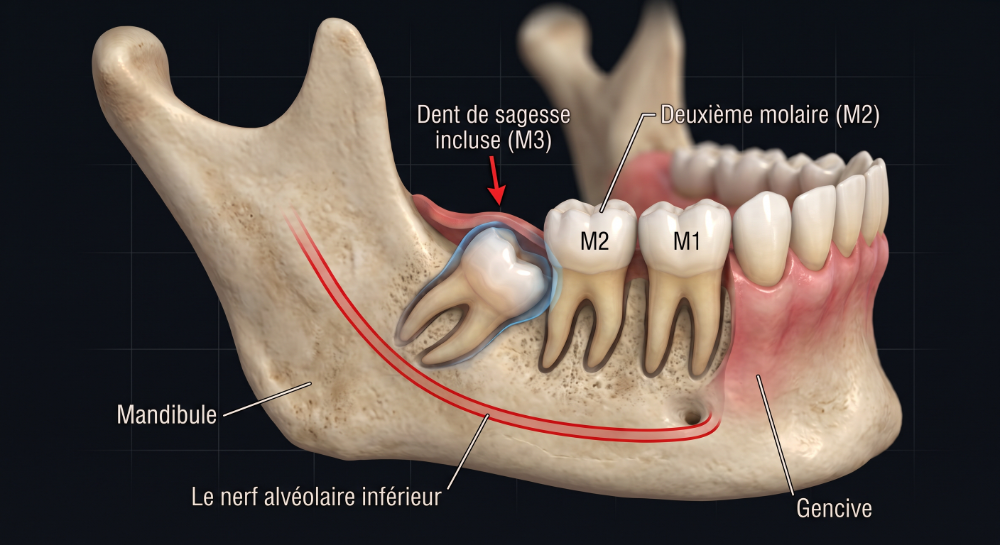
Wisdom teeth are the last teeth to erupt at the back of the dental arch, typically during late adolescence or early adulthood. When there is insufficient space within the dental arch, these teeth may be unable to erupt properly and become trapped in the jawbone against the adjacent second molars. When this occurs, they are classified as impacted wisdom teeth.
Why Is Extraction Necessary?
Extraction of impacted wisdom teeth may be recommended for several clinical reasons:
- Recurrent or Severe Infection (Pericoronitis): An infection of the gum tissue (mucosa) partially overlying the crown of an impacted tooth. Symptoms include localized pain, gingival swelling, facial swelling, and restricted mouth opening (trismus).
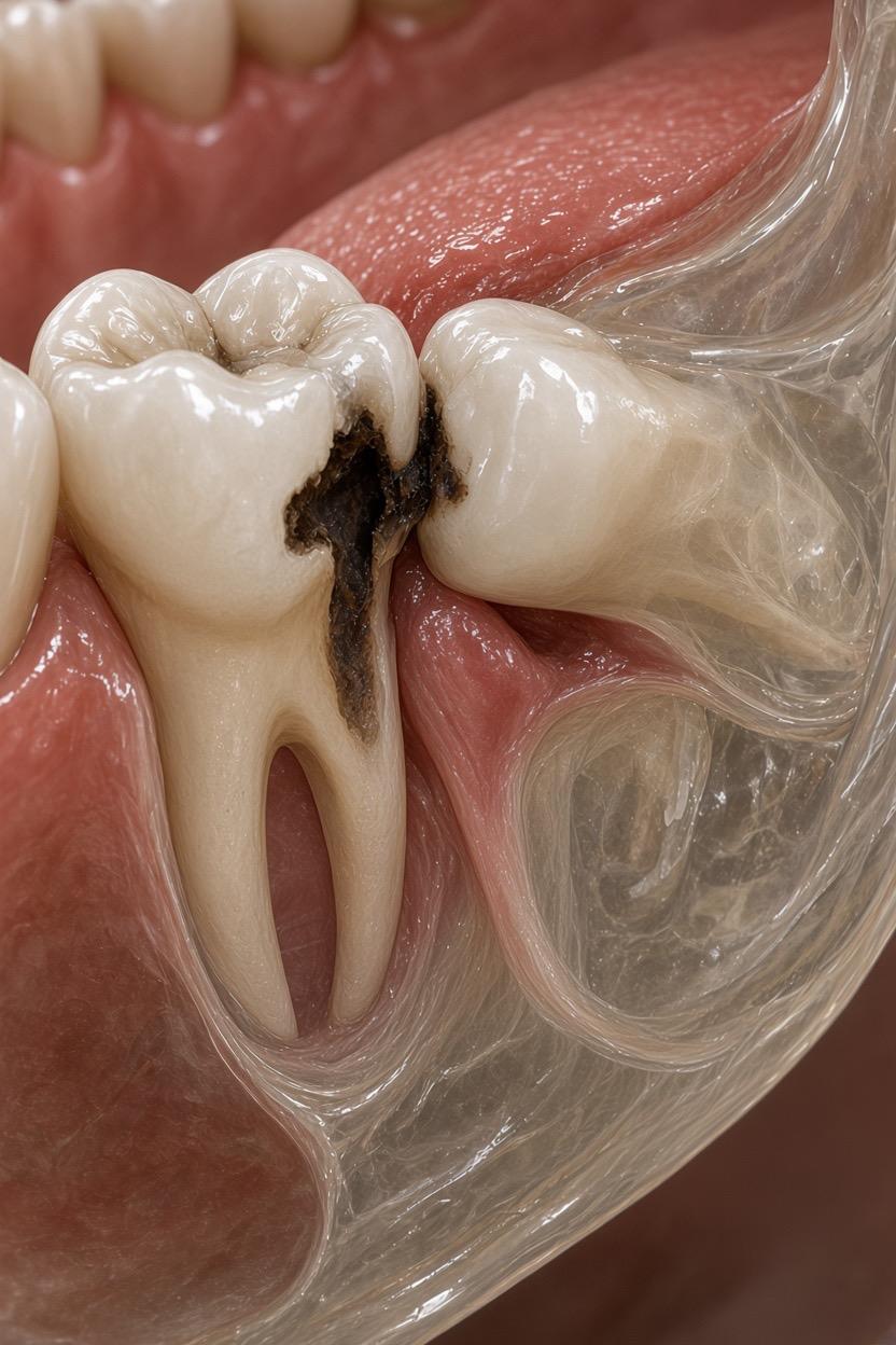
- Dental Caries: Tooth decay affecting the impacted wisdom tooth or the inaccessible posterior surface of the second molar.
- Soft Tissue Irritation: Malpositioned wisdom teeth that erupt buccally (toward the cheek), leading to recurrent mechanical trauma, mucosal ulceration, and inflammation.
- Orthognathic Surgery Preparation: Prophylactic removal prior to planned surgical jaw advancement or setback procedures to minimize complication risks and facilitate osteotomy sites.
- Periodontal Disease: Food impaction and poor hygiene accessibility causing chronic periodontal pocketing and bone loss around the second molars.
- Cystic Lesions: Formation of odontogenic cysts (e.g., dentigerous cysts) associated with the follicle of the impacted tooth.
- Associated Neoplasms: Rare development of benign or malignant odontogenic tumors.
- Orthodontic Considerations: Space creation required by your dentist or orthodontist to align teeth, prevent late crowding, or minimize relapse post-orthodontic therapy.
- Mandibular Fractures: An impacted tooth situated along a jaw fracture line that must be removed to facilitate anatomical reduction and proper fixation.
Are There Alternatives to Extraction?
Conservative management includes analgesics and anti-inflammatories (e.g., paracetamol, ibuprofen) to control acute pain and swelling, as well as oral antibiotics and antiseptic mouth rinses (chlorhexidine or povidone-iodine / Isobetadine) for active infection. However, these are strictly symptomatic, temporary measures that do not address the underlying anatomical problem.
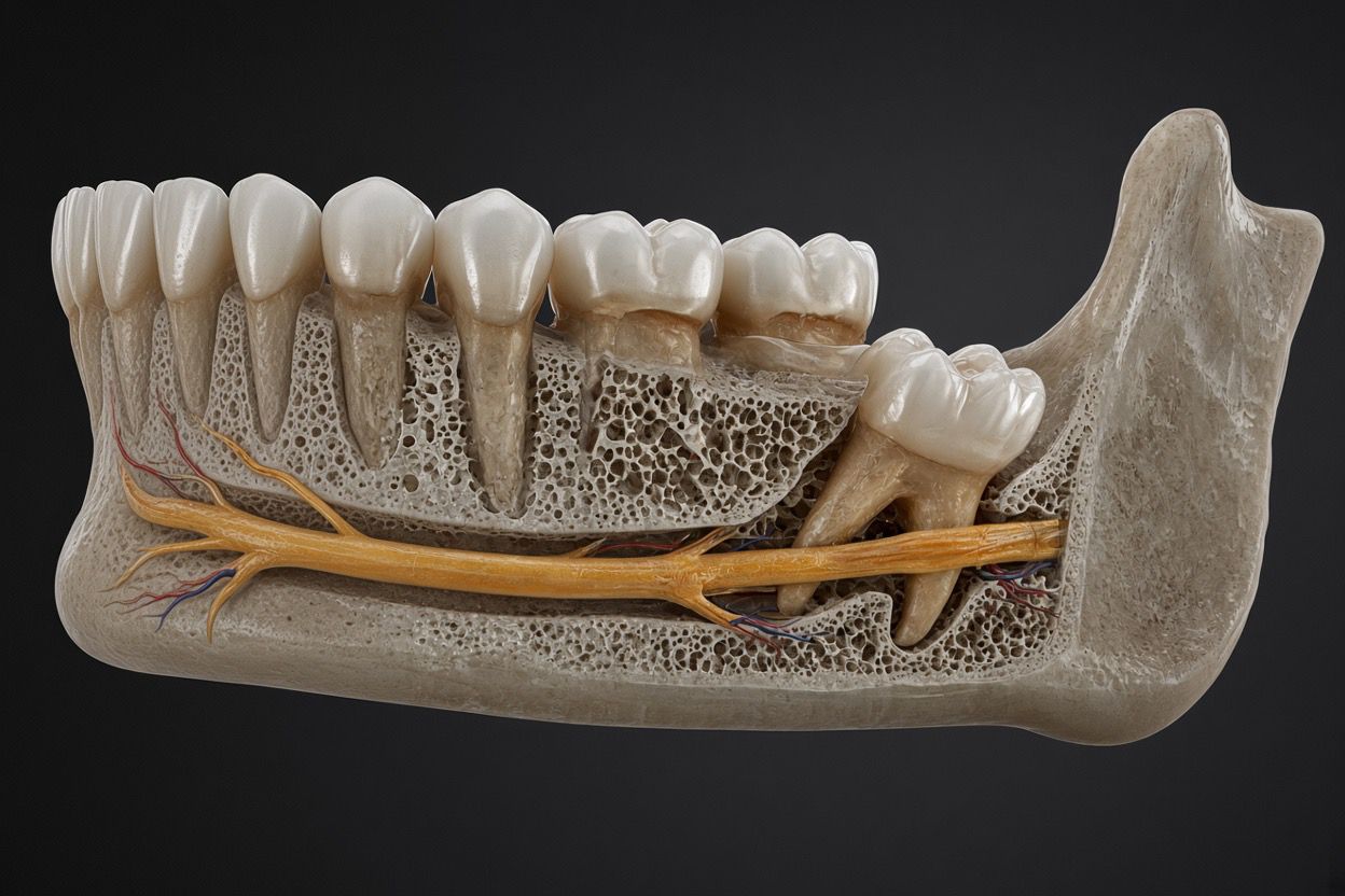
Coronectomy: If radiographic imaging demonstrates that the root apex lies in immediate proximity to the inferior alveolar nerve, a coronectomy may be considered. In this procedure, only the coronal crown of the impacted tooth is removed, leaving the roots intentionally undisturbed in the bone to drastically reduce the risk of permanent nerve injury (paresthesia).
The Surgical Procedure
The procedure may be conducted under local anesthesia, intravenous sedation, or general anesthesia. Dr. Yousefpour will review the surgical plan and advise on the most suitable anesthetic technique depending on anatomical complexity, the number of teeth requiring extraction, and patient preference.
Key procedural aspects include:
- Duration: Typically ranges between 30 and 90 minutes depending on difficulty.
- Surgical Access: An incision is made along the oral mucosa to elevate a mucoperiosteal flap and expose the underlying bone.
- Bone Removal and Sectioning: A small amount of overlying cortical bone is gently removed. If necessary, the crown and roots are sectioned into smaller segments to enable atraumatic removal.
- Wound Closure: The surgical site is cleaned, irrigated, and closed with absorbable sutures, which spontaneously dissolve within 3 weeks.
Potential Complications & Management
| Complication | Clinical Description | Preventative & Management Strategy |
|---|---|---|
| Postoperative Pain | Variable discomfort corresponding to procedural duration and degree of bone removal. | Apply cold packs during the first 24 hours. Take prescribed analgesics and anti-inflammatory medications. |
| Bleeding / Hemorrhage | Mild oozing expected from the surgical socket within the first 24 hours. | Apply direct bite pressure over sterile gauze for 30 minutes. Avoid spitting, rinsing, and hot foods/liquids for 24 hours. |
| Swelling and Bruising | Expected inflammatory reaction, particularly pronounced with lower wisdom teeth. | Intermittent cold pack therapy, head elevation during rest, and adherence to anti-inflammatory regimens. |
| Postoperative Infection | Swelling or pain that worsens past postoperative day 4 or 5, potentially with palpable swelling or purulent discharge. | Clinical evaluation by Dr. Yousefpour, culture if necessary, and targeted antibiotic therapy. |
| Dry Socket (Alveolar Osteitis) | Premature breakdown or dislodgment of the primary blood clot, leaving exposed alveolar bone; presents with severe radiating pain around day 2 to 3. | Strict avoidance of smoking, vigorous rinsing, or drinking with straws. Managed via socket irrigation and medicated soothing dressings. |
| Nerve Injury (Paresthesia) | Altered sensation, numbness, or tingling in the lower lip or chin due to proximity to the inferior alveolar nerve. | Usually temporary, rarely permanent. In high-risk cases, coronectomy or intentional retention of small root tips is performed. |
| Adjacent Tooth Damage | Fracture, decementation of crowns, or loss of existing restoration on the adjacent second molar. | Careful elevation technique and pre-extraction assessment of adjacent restorations. |
| Oroantral Communication | Perforation between the maxillary sinus cavity and oral cavity during upper molar extraction. | Sinus precautions (no nose blowing), antibiotic coverage; surgical flap closure if required. |
| Sinus Displacement | Accidental displacement of root tip or crown into the maxillary antrum. | Specialized surgical retrieval (Caldwell-Luc or transoral endoscopic approach). |
| Mandibular Fracture | Very rare mechanical failure during deep impaction removal in dense bone. | Open or closed reduction with rigid fixation (plates and screws). |
| Trismus (Limited Opening) | Spasm or inflammation of the masticatory muscles, frequent after lower extractions. | Gentle jaw-opening exercises, moist heat after 48 hours, and anti-inflammatories. Gradual recovery over 1–3 weeks. |
Postoperative Care & Recovery Instructions
Adherence to postoperative guidelines promotes optimal healing and reduces complication risks:
- Immediate Recovery (General Anesthesia): Patients are monitored in the post-anesthesia care unit before discharge. A responsible adult companion must escort you home and remain with you for at least 24 hours.
- Hemostasis: If minor bleeding resumes, place a sterile gauze directly over the extraction site and bite firmly for 10 to 30 minutes without disturbance.
- Medication Compliance: Take all prescribed medications as instructed — analgesic (paracetamol/Dafalgan) for pain relief, anti-inflammatory (ibuprofen) for swelling, antibacterial rinse (chlorhexidine/Corsodyl or povidone-iodine/Isobetadine) starting 24 hours post-surgery, and the full course of oral antibiotics if prescribed.
- Dietary Adjustments: Maintain a soft, cool, or room-temperature semi-liquid diet for several days until chewing is comfortable. Avoid hot, spicy, crunchy, or brittle foods.
- Activity & Rest: Avoid vigorous physical exercise, heavy lifting, or bending over during the acute recovery period. Plan for roughly one full week off work or academic duties to rest. In the absence of complications, standard activities can typically be resumed after 7 days.
- Oral Hygiene & Habits: Strictly avoid smoking and alcohol intake. Brush remaining teeth carefully, avoiding direct trauma to healing surgical wounds.
Visual Reference: Impacted Wisdom Teeth
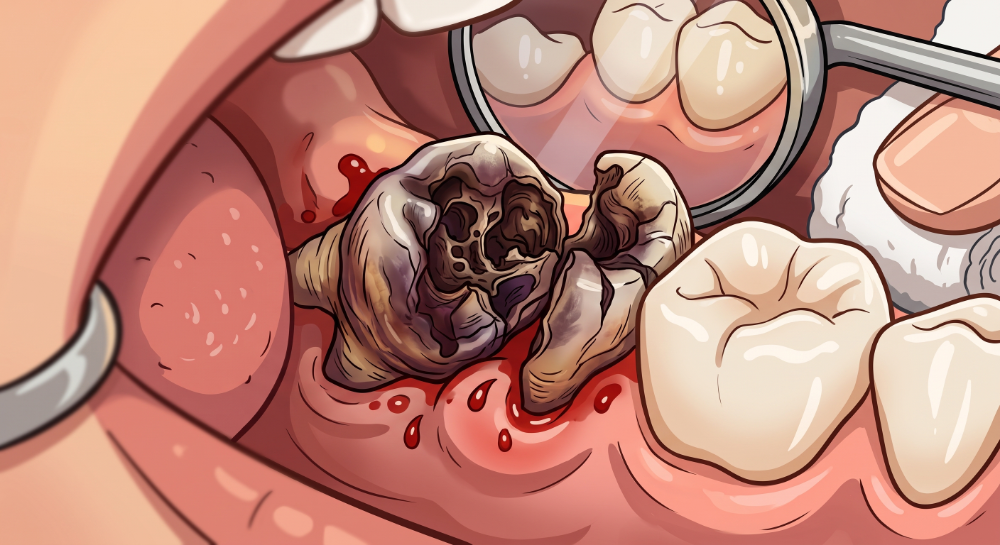
A visual summary of impacted wisdom tooth anatomy, decay progression, and clinical presentation.