



Maxillary Sinus Lift
Dr. Yousefpour — Maxillofacial Surgeon & Stomatologist
Specialized expertise in reconstructive bone surgery
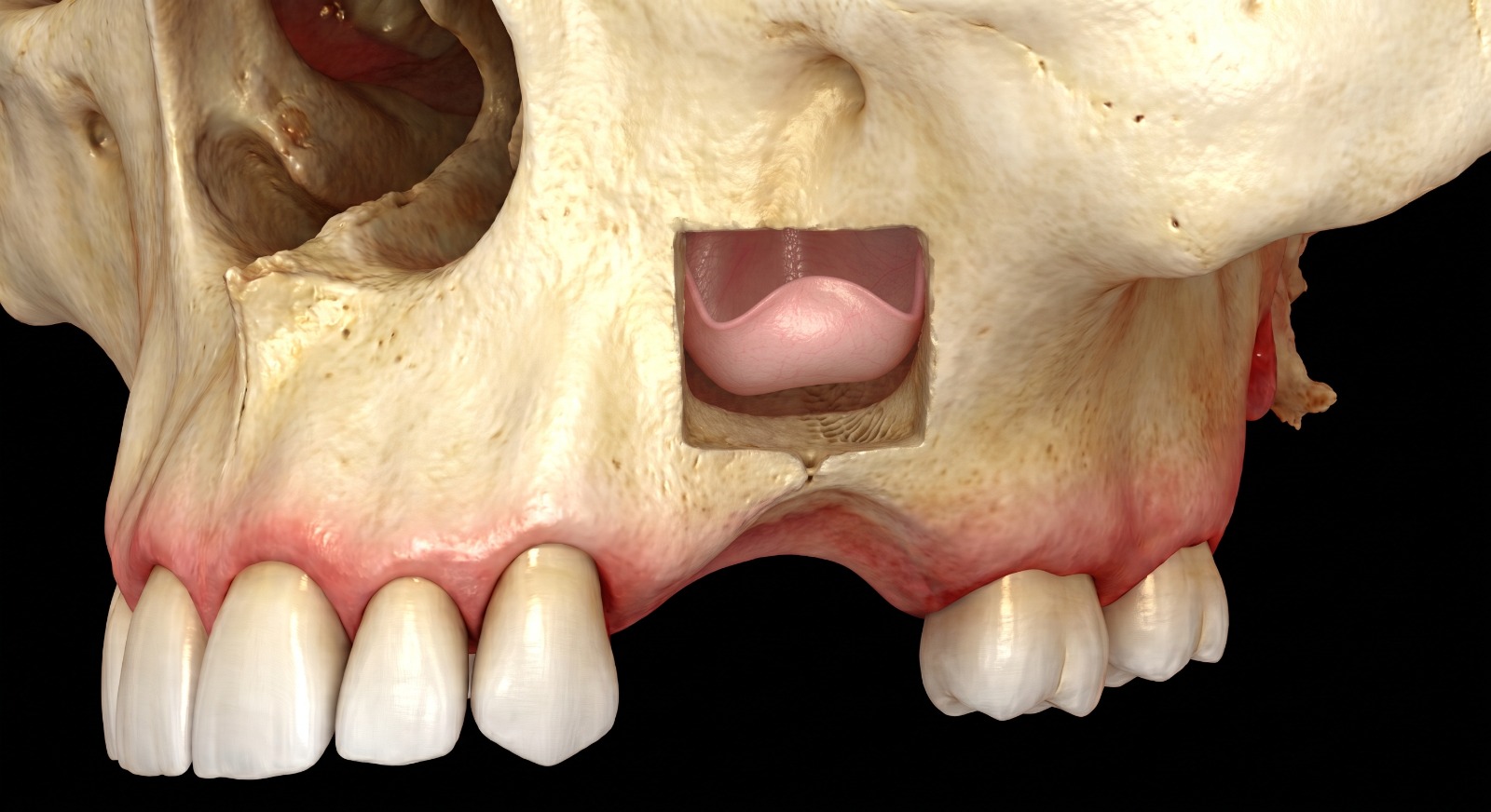
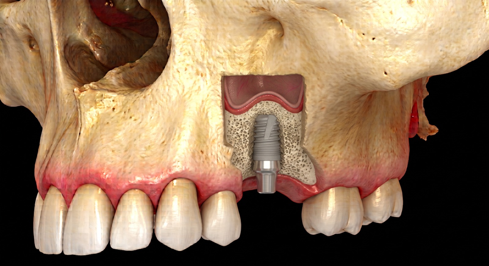
The loss of upper premolars or molars inevitably leads to a loss of bone height, often accentuated by the natural expansion of the maxillary sinus (sinus pneumatization).
When there is not enough bone volume to anchor a dental implant stably and durably, the sinus lift (or elevation of the sinus floor) is the procedure of choice. As a maxillofacial surgeon, Dr. Yousefpour brings in-depth mastery of sinus anatomy, the airways, and advanced grafting techniques to ensure a safe, predictable, and comfortable procedure.
Why Trust Dr Yousefpour With Your Sinus Lift?
- Complete anatomical & ENT mastery: A comprehensive assessment of sinus patency, the middle meatus, and anatomical variations (Underwood's septa).
- Precision digital technology: Advanced 3D planning using Cone Beam CT (CBCT) and guided surgery for millimeter precision.
- Surgical safety & comfort: Performed under advanced local anesthesia or sedation, in a dedicated sterile surgical environment.
Two Surgical Approaches, Matched to Your Situation
The choice of technique depends directly on the initial bone thickness observed on your 3D assessment:
| Surgical Approach | Clinical Indication | Protocol & Implant Placement |
|---|---|---|
| Crestal Approach (Minimally Invasive) | Moderate deficit (bone height > 5mm) | Direct access through the implant drilling site. The implant is almost always placed during the same session. |
| Lateral Approach | Severe deficit (residual height < 5mm) | Creation of a small lateral window to graft a larger volume. Implant placement is either simultaneous or delayed, depending on primary stability. |
Surgical Protocol & Technological Innovations
Dr. Yousefpour integrates the most modern regenerative protocols to optimize healing and minimize post-operative effects:
- Piezoelectric osteotomy (ultrasound): Selective bone cutting using micro-vibrations, drastically reducing the risk of injury or perforation of the Schneiderian membrane.
- Certified augmentation biomaterials: Use of autologous bone (harvested locally) and/or high-purity biocompatible bone substitutes to induce dense new bone formation.
- Platelet concentrates (PRF): Optional use of autologous growth factors to accelerate vascularization and tissue regeneration.
- Resorbable barrier membranes: Optimal protection of the grafted site with a collagen membrane to guide bone regeneration.
Your Care Pathway
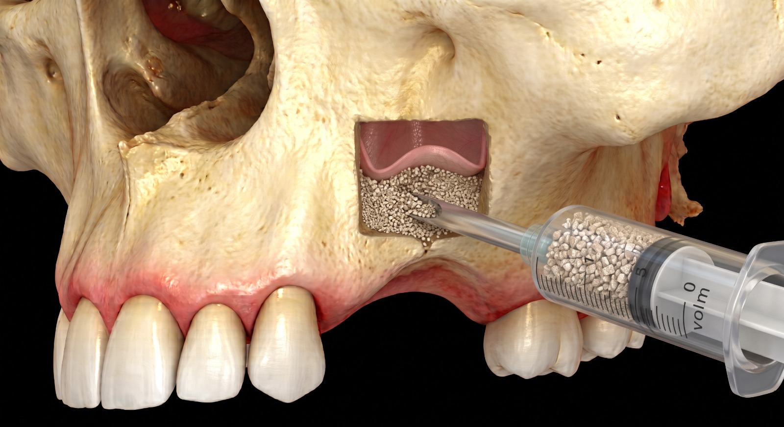
- Consultation & 3D assessment: CBCT scan analysis, implant feasibility study, and a tailor-made treatment plan.
- Surgical stage: A procedure performed with complete peace of mind under an anticipated pain-management protocol (duration: 45 to 75 minutes).
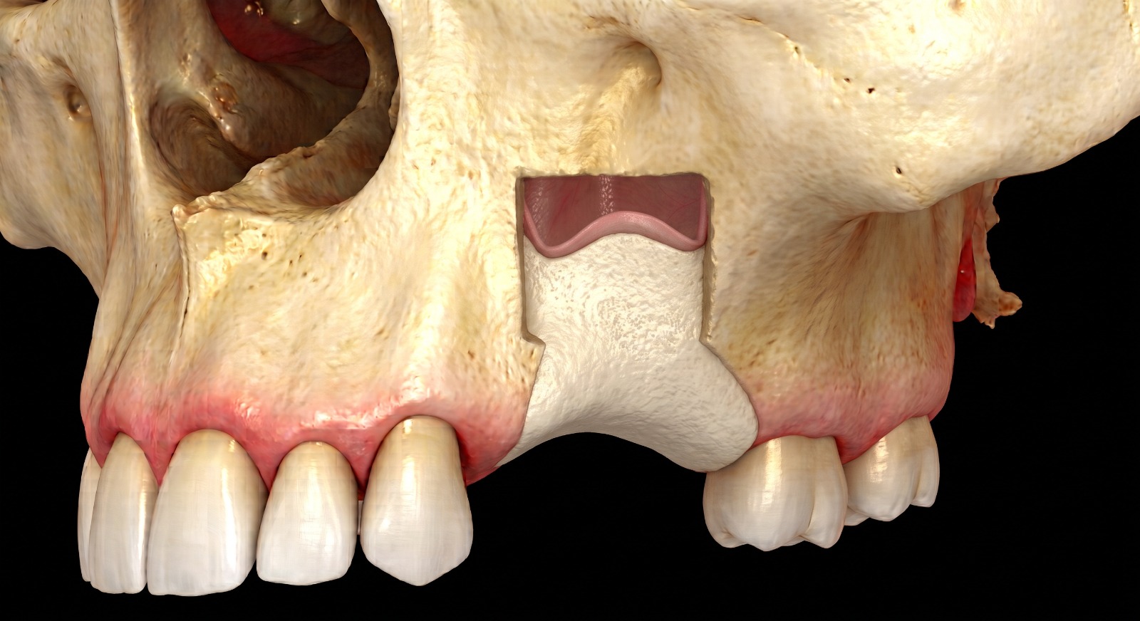
- Maturation phase: A natural osseointegration period of 4 to 6 months, transforming the biomaterial into solid, living bone.
- Implant-prosthetic finalization: Placement of the final crown by your regular dentist, on a perfectly stable bone foundation.
Recommendations & Post-Operative Follow-Up
Dr. Yousefpour provides rigorous, personalized follow-up. Post-operative effects today are very manageable thanks to minimally invasive techniques:
- Immediate advice: Avoid forceful nose-blowing, sneeze with your mouth open, and suspend air travel or intense sports activity for 10 to 14 days.
- Pain management: Targeted prescription of pain relievers and appropriate local care for a fast, comfortable recovery.
Not Enough Bone for Implants?
Book a consultation with Dr. Yousefpour for a personalized 3D evaluation and discover the solution best suited to your situation.